I’ve been working on the rendering engine for SynthNet, so both the neuronal morphologies can be seen, and membrane potential and other activity can be visualized through color coding, like in an fMRI.

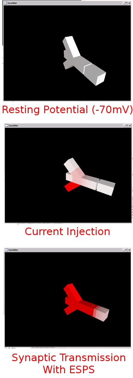
This is an example of it in action so far. The models look pretty clunky and simple, but that’s by design – I just want a simple, lightweight viewer that doesn’t take up too much CPU time. This example shows two neurons and a synapse – one with 3 dendrites and an axon, with a synaptic gap between the post-synaptic neuron, consisting of only a small neuronal segment (a dendrite). Membrane potential is shaded in red, with white representing -70mV, and bright red representing 30mV.
The top picture shows the two neurons at rest. In the second picture, I inject current into one of the dendrites, and you can see the current flowing up its process into neighboring dendrites and the soma, getting weaker as it goes due to attenuation.
In the final picture, we see a large amount of current injection into all dendrites, causing an action potential and release of neurotransmitters into the synaptic gap, which causes an influx of ions in the post-synaptic neuron, depolarizing the membrane potential, as can be see with the post-synaptic neuron also showing red shading.
When I first got the code working and watched all this happening live, it was definitely one of those wow moments.
 Hello - and thanks for visiting my site! I maintain ToniWestbrook.com to share information and projects with others with a passion for applying computer science in creative ways. Let's make the world a better and more beautiful place through computing! | More about Toni »


Leave a Reply